info@harmanismed.gr

Anatomy Models
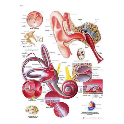
Ear anatomy map
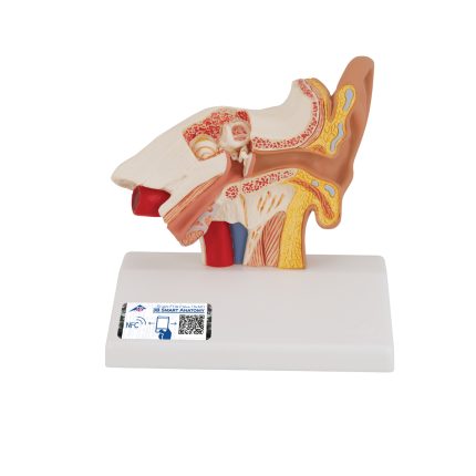
Ear model 1000252
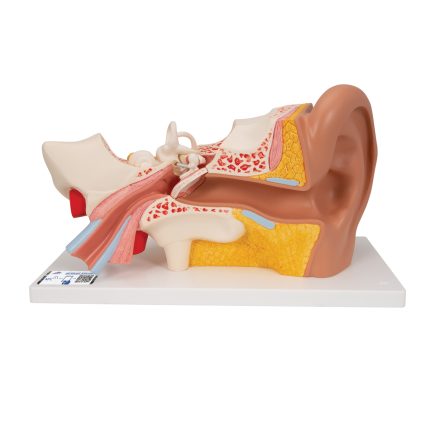
Ear model, 3 times life size, 4 parts 1000259
Ear model, 3 times life-size, 4-part 1000259 representing the outer, middle and inner ear. The detailed human ear model features a removable eardrum with malleus, anvil and stirrup, as well as a 2-part labyrinth with cochlea and auditory/balancing nerve. The ear is on a stand for easy display in the classroom or doctor's office. This ear model is a great way to teach and study the anatomy of the human ear! Excellent model for teaching-learning the anatomy of the human eye! Human eye on a stand. It has an app for your smartphone, tablet or desktop device.
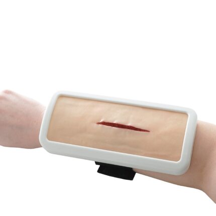
Educational model of sutures in the form of a cushion H130621
The H130621 suture training model has the shape of a rectangular pillow and allows you to learn surgical suturing and knotting techniques under realistic conditions.
- The H130621 suture training model consists of special foam and "artificial leather" made of soft PVC.
- A special, soft PVC was chosen for the H130621 suture training model, which very naturally reflects the properties of real skin and allows the user to practice skin suturing and surgical knot techniques under realistic conditions.
- In addition, special replacement leather pads are available, which means that they can be economically replaced.
- It features a pre-cut wound that can be stitched over and over again. If necessary, additional wounds can be added.
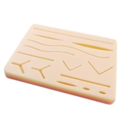
Educational Skin Stitching Template in Various Designs
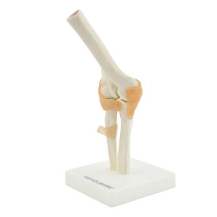
Elbow Joint Model - 130492

Elbow joint model with ligaments 1000165
Elbow Joint Model with Ligaments 1000165 provides a graphic demonstration of the anatomy and mechanics of the human elbow joint. Use this fully flexible model to demonstrate abduction, anterior rotation, posterior rotation, and internal/external rotation. The elbow joint consists of a portion of the humerus, complete ulna and radius, and joint ligaments. Comes on a removable base for easy study or demonstration. Includes an app for your smartphone, tablet, or desktop device.
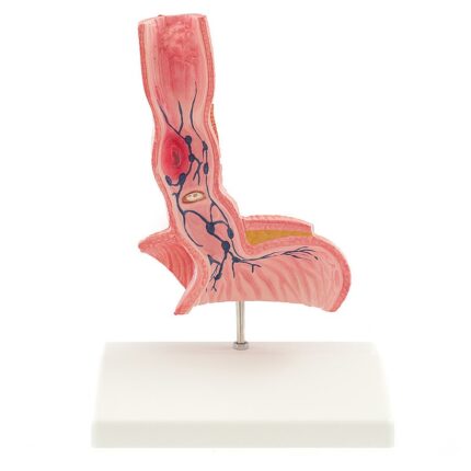
Esophageal model 139049
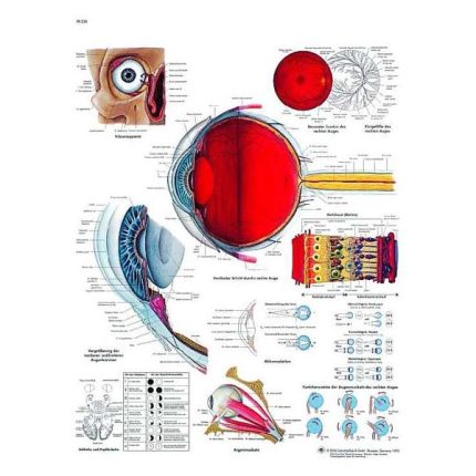
Eye anatomy map
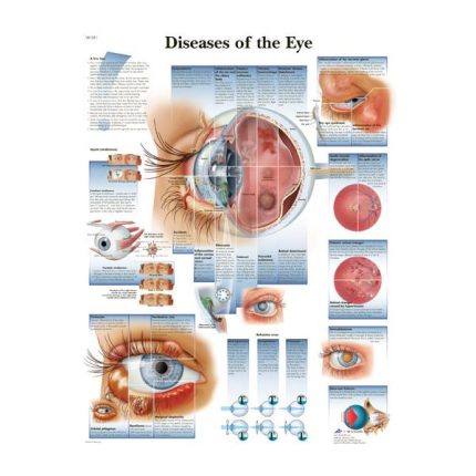
Eye disease anatomy map
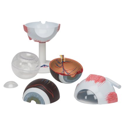
Eye model, 3 times full size, 6 parts 1000259
Eye model, 3 times full size, 6 parts 1000259 which breaks down into:
- Two halves of the sclera with corneal and ocular muscle attachments
- Two halves of the choroid with iris and retina
- Eye lens
- Vitreous body
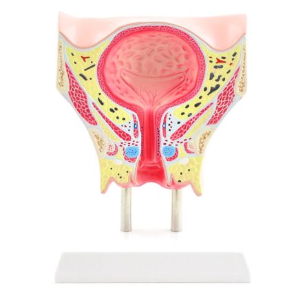
Female bladder model H130481
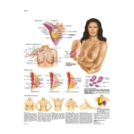
Female breast anatomy chart
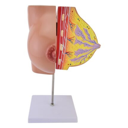
Female Breast Model 139032
Female breast model of Heine Scientific made in Germany. This breast model from HeineScientific shows the anatomical structures of the female breast. Both parts of the anatomical model are connected by a hinge so that the chest can be opened. This allows you to easily see internal structures such as the milk ducts, mammary glands, and adipose tissue. Also suitable as a teaching tool for schools and universities
It has a base.
It has a base.
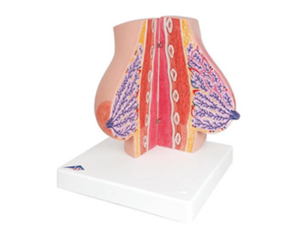
Female Breast Model 3B
A model of women's breasts of the company 3B made in Germany with a size of 37 cm. Top quality with medically correct breast anatomy makes the product a great educational tool for you. The models are held in place with magnets for easier display. Right breast milk:
Divided into 2 parts, held in place by magnets
Healthy breast milk tissue on the "cut" surface of the outer half
Inflammation of the breast gland (mastitis) on the "cut" surface of the inner half
3 years warranty
Divided into 2 parts, held in place by magnets
Healthy breast milk tissue on the "cut" surface of the outer half
Inflammation of the breast gland (mastitis) on the "cut" surface of the inner half
3 years warranty
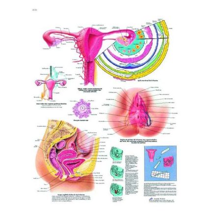
Female genital anatomy chart
Female genital anatomy chart with metal slat
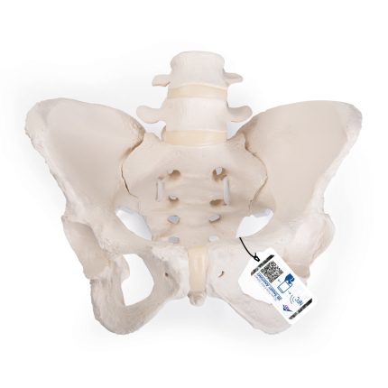
Female pelvic skeleton model 1019864
Female Pelvis Skeleton Model 1019864 "Bungee" made in a natural way. All pelvic bones are flexibly positioned and can be easily disassembled. Available with heads without femur, these female pelvis models perfectly demonstrate the pelvic joints. Particularly suitable for patient education, as well as a useful learning tool for midwives and in the fields of Obstetrics, Gynecology and Orthopedics.
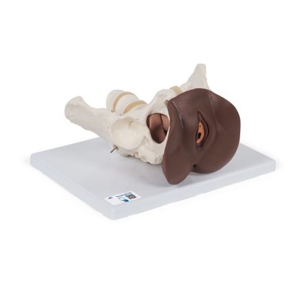
Female pelvic skeleton model with genitals, 3 parts 1024386
Female pelvic skeleton with genitals 3-part model 1024386 which is particularly suitable for studying the female genitals in the context of their anatomical position in the pelvis. It consists of a female pelvis with movable symphysis, ischium, sacrum, coccyx, 2 lumbar vertebrae and a female genital insert with rectum. The uterus and bladder can be removed for more detailed anatomical study. This model is ideal for studying and teaching the anatomy of the female genitals. The female pelvic skeleton with genitals is delivered on a base for easy display in the classroom or in the doctor's office.
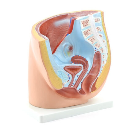
Female Pelvis Model 130380
Model of a female pelvis by Heine Scientific. This pelvic model shows the anatomical structures of an adult woman's pelvis in an obelisk incision. Thanks to the multitude of anatomical structures represented, the female pelvic model is suitable for use in gynecology, as well as urology. It is suitable for biology courses, as well as for explaining diseases, operations or anatomical features to patients. This female pelvic model is just as popular in gynecological practice as it is among midwives. In order to better highlight the various anatomical structures, the female pelvic model has been colored accordingly. It is three-dimensional and comes mounted on a base.
Dimensions: 25 x 18 x 28 cm (D x W x H)
Dimensions: 25 x 18 x 28 cm (D x W x H)
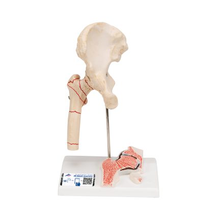
Femur fracture and hip osteoarthritis model 1000175
Femur fracture and hip osteoarthritis model 1000175 was developed to provide patients with understandable information, e.g. before surgery. It shows the right hip joint of an elderly person in half life size. In addition to the external anatomy of the hip joint, a frontal section through the femoral neck is depicted in relief on the base. The hip joint model shows the most common femur fractures in practice, as well as the typical symptoms of wear and tear of the hip joint (hip arthritis or hip osteoarthritis). It has an app for your smartphone, tablet or desktop device.
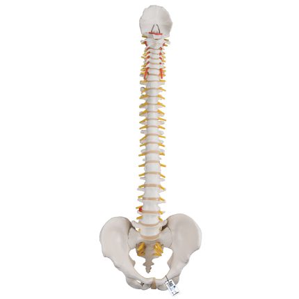
Flexible spine model 1000121
Flexible Spine Model 1000121 for patient and student education. This spine is fully flexible and designed for hands-on demonstrations. The spine includes the following features:
- Complete male pelvis and occipital plate
- Fully flexible placement on the spine
- L3-L4 herniated disc in the spine
- Spinal nerve exits
- Cervical vertebral artery
- This high-quality spine is an excellent tool for teaching/learning the anatomy of the human spine.
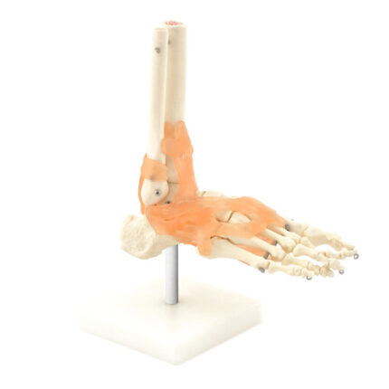
Foot - ankle model 130247
Full-size model of foot and ankle demonstrates the structure and movement of the human foot with all the bones and the most important ligaments. It is mounted on a metal support, allowing the rest of the foot to move in different positions. For a more complete performance it includes the ankle and the lower parts of the fibula and tibia.
Dimensions: approx. 20 x 20 x 8 cm (W x H x W)
Dimensions: approx. 20 x 20 x 8 cm (W x H x W)
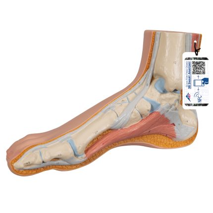
Foot model 1000354
Foot model 1000354 where the superficial structures of the foot are presented in this high-quality model. The internal bones, muscles, ligaments and nerves are also represented in this normal foot skeleton. The osteoporosis model is an excellent high-quality anatomical teaching aid. It has an app for your smartphone, tablet or desktop device.