info@harmanismed.gr

3B scientific
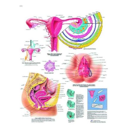
Female genital anatomy chart with metal slat
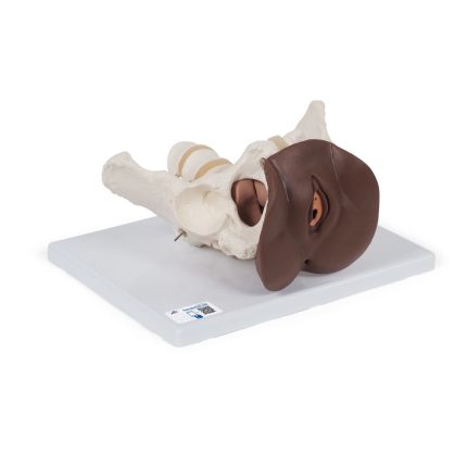
Female pelvic skeleton model with genitals, 3 parts 1024386
Female pelvic skeleton with genitals 3-part model 1024386 which is particularly suitable for studying the female genitals in the context of their anatomical position in the pelvis. It consists of a female pelvis with movable symphysis, ischium, sacrum, coccyx, 2 lumbar vertebrae and a female genital insert with rectum. The uterus and bladder can be removed for more detailed anatomical study. This model is ideal for studying and teaching the anatomy of the female genitals. The female pelvic skeleton with genitals is delivered on a base for easy display in the classroom or in the doctor's office.
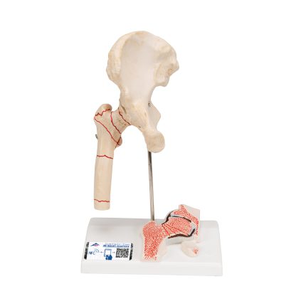
Femur fracture and hip osteoarthritis model 1000175
Femur fracture and hip osteoarthritis model 1000175 was developed to provide patients with understandable information, e.g. before surgery. It shows the right hip joint of an elderly person in half life size. In addition to the external anatomy of the hip joint, a frontal section through the femoral neck is depicted in relief on the base. The hip joint model shows the most common femur fractures in practice, as well as the typical symptoms of wear and tear of the hip joint (hip arthritis or hip osteoarthritis). It has an app for your smartphone, tablet or desktop device.
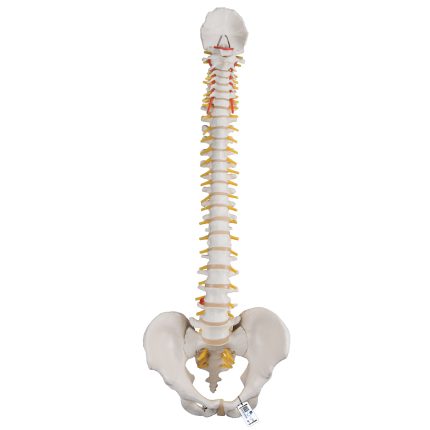
Flexible spine model 1000121
Flexible Spine Model 1000121 for patient and student education. This spine is fully flexible and designed for hands-on demonstrations. The spine includes the following features:
- Complete male pelvis and occipital plate
- Fully flexible placement on the spine
- L3-L4 herniated disc in the spine
- Spinal nerve exits
- Cervical vertebral artery
- This high-quality spine is an excellent tool for teaching/learning the anatomy of the human spine.
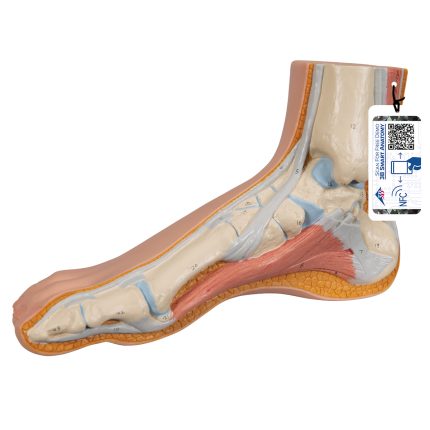
Foot model 1000354
Foot model 1000354 where the superficial structures of the foot are presented in this high-quality model. The internal bones, muscles, ligaments and nerves are also represented in this normal foot skeleton. The osteoporosis model is an excellent high-quality anatomical teaching aid. It has an app for your smartphone, tablet or desktop device.
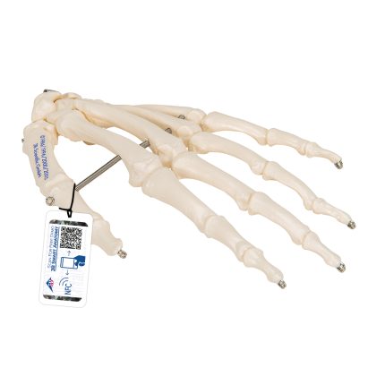
Hand model on wire 1019367
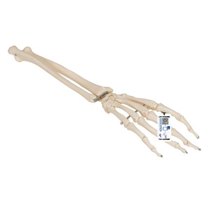
Hand skeleton model with ulna and radius 1019370
Hand skeleton model with ulna and radius 1019370 which is fixed with wire, naturally flexible through the wrist. It is slightly flexible through the fingers to simulate the natural movement of the hand. Unfortunately, it is not possible to specify the left or right version, you will receive a left or right side model at random. It has an app for your smartphone, tablet or desktop device.
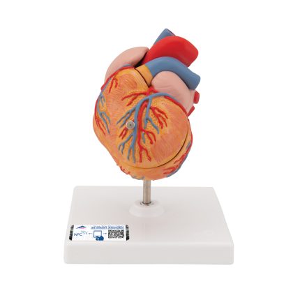
Heart model with left ventricular hypertrophy, 2 parts 1000261
2-part heart model with left ventricular hypertrophy 1000261 showing the long-term effects of increased cardiac activity due to high blood pressure. The muscular wall of the left heart ventricle is significantly thicker and the edge of the heart is visibly rounded. Highly detailed 2-part heart where the front wall of the heart is removable to reveal the chambers and valves inside. The heart is slightly smaller than life-size with excellent anatomical detail throughout. Heart on a removable base. Features an app for your smartphone, tablet or desktop device.
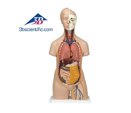
Human body model 3B 90cm with 12-parts
3B human body model made in Germany, 90 cm in size, consisting of 12 parts. Top quality with medically correct anatomy with a unisex torso hand-painted in detail and made of high quality plastic. The human body model is disassembled into 12 parts and is ideal for clinics, schools and medical students.
Dimensions: 87 x 38 x 25 cm
Weight: 4.6 kg
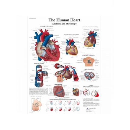
Human Heart Anatomy Map
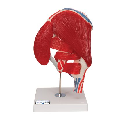
Human Hip Joint Model with Removable Muscles 7 part 1000177
Hip joint model with removable muscles 7 parts 1000177 depicting the right hip joint of a man with the individual muscles, as well as the origins and insertions of the muscles on the femur and hip bone. For educational purposes, the areas of origin and insertion of the muscles have been raised and shown in color (muscle origin = red, muscle insertion = blue) on the hip joint. The hip muscles have been placed in the corresponding areas of origin and insertion and are removable from the hip joint for a different perspective. Studying the anatomy of the hip bone is easy with this hip joint and removable muscle. The relationship between the muscular and skeletal systems is clearly represented with this hip joint model. It has an app for your smartphone, tablet or desktop device.
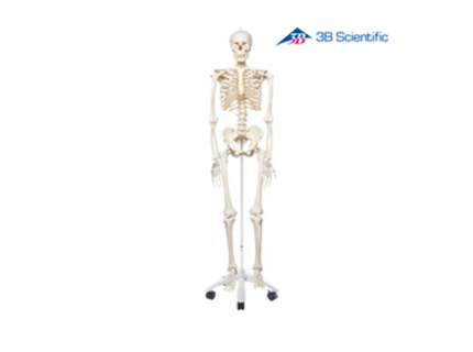
Human skeleton wheeled 3B - A10
Human skeleton on wheels by 3B, made in Germany. It is an accurate anatomical skeleton model of excellent quality suitable for use in hospitals, schools, universities, as well as for laboratories. It has excellent quality of construction and materials, is medically accurate and has a base with 5 wheels and a cover case. Made of durable, unbreakable plastic material.
3 year warranty

Kidney anatomy map
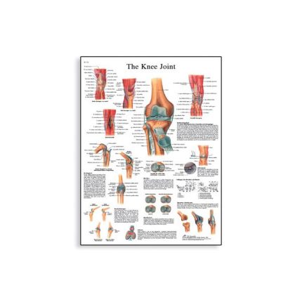
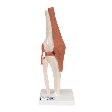
Knee joint model with ligaments 1000163
Knee Joint Model with Ligaments 1000163 accurately demonstrates the structure and movement of the knee joint. Designed for anatomical education and clinical demonstration, the model provides a clear understanding of the knee's biomechanics and joint function. Mounted on a removable base, the model is suitable for both classroom teaching and close-up anatomical study. It has an app for your smartphone, tablet or desktop device.
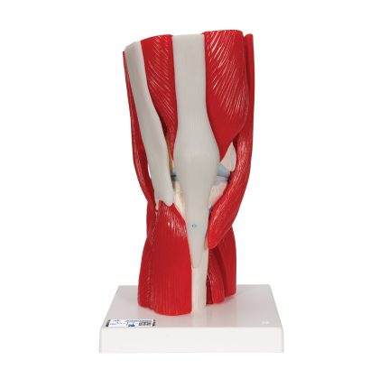
Knee joint model with removable muscles, 12 parts 1000178
The 12-part knee joint model with removable muscles 1000178 is part of a series of high-quality models. It has been manufactured with precise anatomical accuracy with realistic colors of the joint, bones and muscles. Extremely durable, non-hazardous material of the highest standards has been used throughout the production process. The model consists of 12 parts that can be easily disassembled for easy demonstration. The muscles and muscle parts can be removed and special color coding has been used to show the origin and insertion points of the muscles on the femur, tibia and fibula. In addition, it also shows parts of the peroneal and tibial lateral ligaments. Whether you are studying or teaching the deeper anatomical layers of the human knee joint, or looking for a tool to better educate your patients about injuries and rehabilitation, the knee joint model is an excellent teaching tool and offers excellent value. It has an app for your smartphone, tablet, or desktop device.
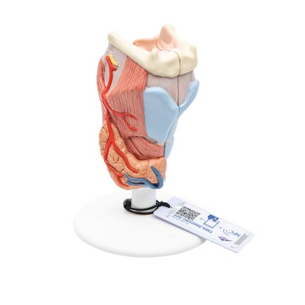
Larynx model 2 parts 1000273
The larynx model is divided into two halves. This larynx model with internal section shows:
- Larynx
- Hyoid bone
- Trachea
- Liaisons
- Muscles
- Vessels
- Nerves
- Thyroid gland
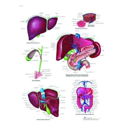
Liver anatomy map
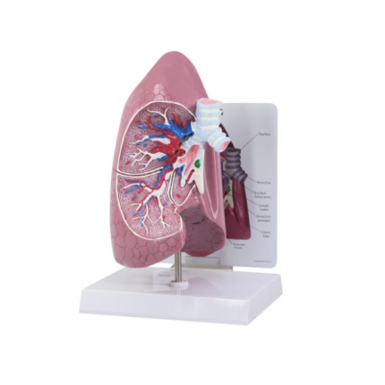
Lung model 1019545
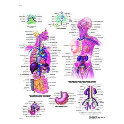
Lymphatic system anatomy map
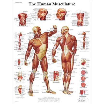
Muscle System Anatomy Map
Anatomy map with dimensions of 50 x 67 cm. The maps are distinguished by their scientific accuracy and are printed on high-quality glossy paper. The map is in English or Latin terminology. Every part of the muscular system is clearly marked. Ideal aid for studying, education, teaching and decorating doctors' offices.
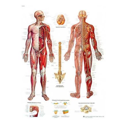
Nervous system anatomy map
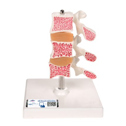
Osteoporosis model 1000153
Osteoporosis model 1000153 consisting of 3 lumbar vertebrae with intervertebral discs, which can be removed. For anatomical comparison, the upper part shows healthy bone structure, the middle part osteoporotic bone structure and the lower part advanced osteoporotic bone structure with flattened plates, deformation and reduced bone mass. For detailed anatomical study, the vertebrae can be removed from the base. The osteoporosis model is an excellent high-quality anatomical teaching aid. It has an app for your smartphone, tablet or desktop device.