info@harmanismed.gr

Anatomy Models
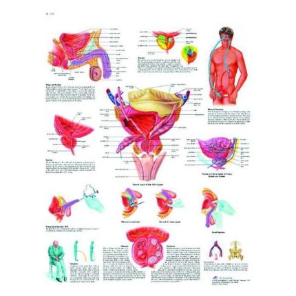
Prostate anatomy map
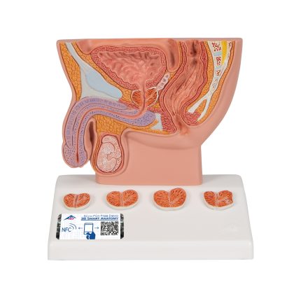
Prostate model 1000319
Prostate model 1000319 provides a cross-section of the male genitalia showing a healthy prostate with bladder, urethra, testicle, symphysis and rectum. Narrowing of the urethra due to prostate changes is made clear by the 4 cross-sections on the base of the prostate model. The anatomical structures of the prostate model have simple schematic coloring. Prostate model on the base. It has an app on your smartphone, tablet or desktop device.

Replacement Lung Bags for Prestan Adult Model
Replacement lung bags for adult model, from Prestan company. Lung bags are used in educational models and simulate the ventilation function of a victim who is not breathing. It would be advisable to replace the lung bags after the end of a training seminar in order to ensure the hygiene of the trainees and prevent the transmission of diseases.
Replacement Lung Bags for Prestan Child Model
Spare lung bags for child model, by Prestan. Lung bags are used in educational models and simulate the ventilation function of a victim who is not breathing. It would be advisable to replace the lung bags after the end of a training seminar in order to ensure the hygiene of the trainees and prevent the transmission of diseases.
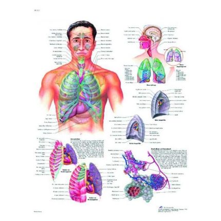
Respiratory system anatomy map

Rheumatoid Disease Anatomy Map
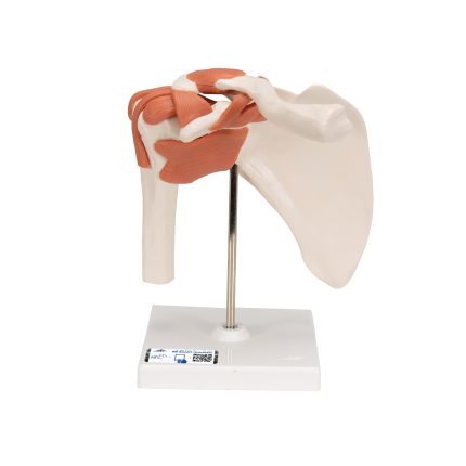
Shoulder joint model 1000159
Shoulder Joint Model 1000159 demonstrates the anatomy and mechanisms of the shoulder joint. Consisting of the scapula, clavicle, humerus, and ligaments of the joint, this fully flexible shoulder joint model clearly demonstrates abduction, anterior rotation, posterior rotation, and internal/external rotation. The shoulder joint model comes on a stand for easy study and display. It has an app for your smartphone, tablet, or desktop device.
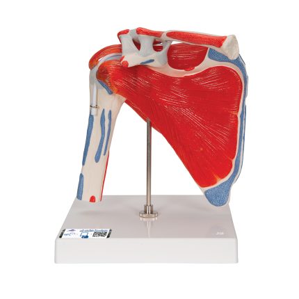
Shoulder joint model with 4 removable muscles, 5 parts 1000176
Shoulder joint model with 4 removable muscles 5 parts 1000176 consisting of the upper half of the humerus, as well as the clavicle and scapula. In addition to illustrating the rotator cuff musculature, the shoulder joint model also shows the origin and attachment points of the shoulder muscles, which are highlighted in color (source = red, attachment points = blue). The shoulder joint model with rotator cuff is mounted on a stand for easy display in the classroom or in the doctor's office. It has an app for your smartphone, tablet or desktop device.
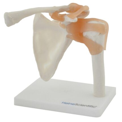
Shoulder Model 130490
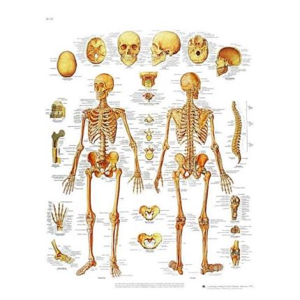
Skeleton anatomy map
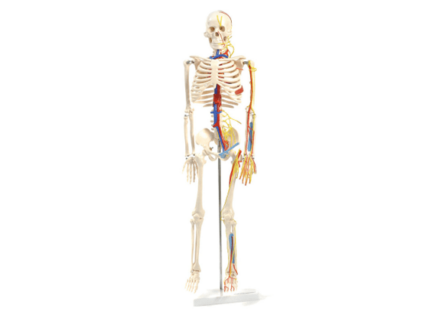
Skeleton with nerves and miniature vessels 130497
Skeleton with nerves and vessels 130497 by Heine Scientific that shows not only the bone structure of the human body, but also several nerve and blood vessel pathways. The skull can be opened to reveal a brain model. To show the circulatory system as fully as possible, a heart is also embedded in this anatomical skeleton.
Vascular details: The vessels are made of special plastic that makes them soft and flexible.
Vascular details: The vessels are made of special plastic that makes them soft and flexible.
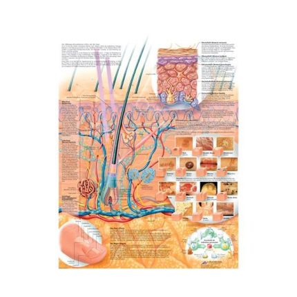
Skin anatomy map
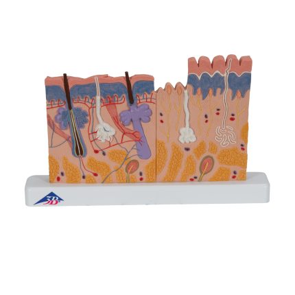
Skin model 1000290
Skin model 1000290 showing the three layers of hairy and glabrous skin, to make the differences between the layers of the skin clear. This skin model features details with hair follicles, sebaceous glands, sweat glands, receptors, nerves and vessels. It is an excellent tool for demonstrating the anatomy of human skin. The skin section is delivered on a base.
The skin section is 40 times larger than full size.
Each original 3B Scientific® anatomy model gives you instant access to its digital twin on your smartphone, tablet or desktop device.
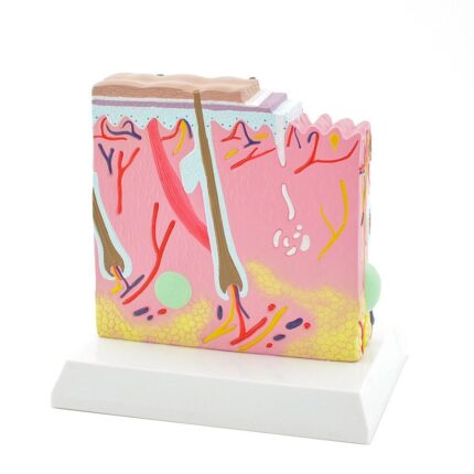
Skin Model 139039
Skin Model by Heine Scientific is a segmental model that clearly shows the structure of the skin, in great detail, from the epidermis to the subcutaneous tissue. It is meticulously painted and is an excellent teaching tool for schools and universities. In addition, it can be used to explain to patients in dermatology clinics or beauty centers. Mounted on a white base.
Dimensions with stand: 20 x 15 x 22.5 cm (H x D x D)
Dimensions with stand: 20 x 15 x 22.5 cm (H x D x D)
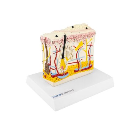
Skin psoriasis model 139058
Skin psoriasis model 139058 showing the normal structure of the skin as well as the effects of psoriasis on the individual layers of the skin.
PRODUCT DETAILS
Block model of skin
Displays effects of psoriasis on various layers of skin
Hand-painted
Fixes firmly on white plastic base
Size (H x W x D): 15.5 x 16 x 11.5 cm
PRODUCT DETAILS
Block model of skin
Displays effects of psoriasis on various layers of skin
Hand-painted
Fixes firmly on white plastic base
Size (H x W x D): 15.5 x 16 x 11.5 cm
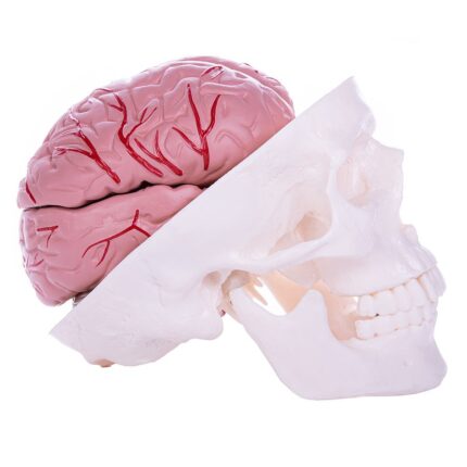
Skull Model With Brain 139150
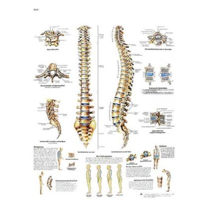
Spinal Anatomy Map
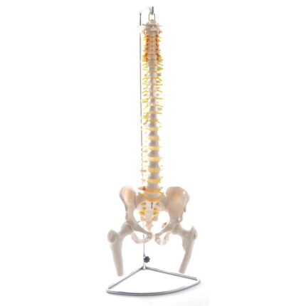
Spinal column model with pelvis and femoral heads 130496
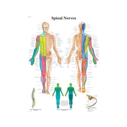
Spinal Nerve Anatomy Map
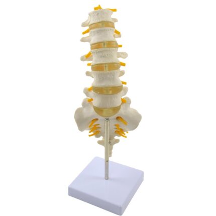
Spine model - 131069
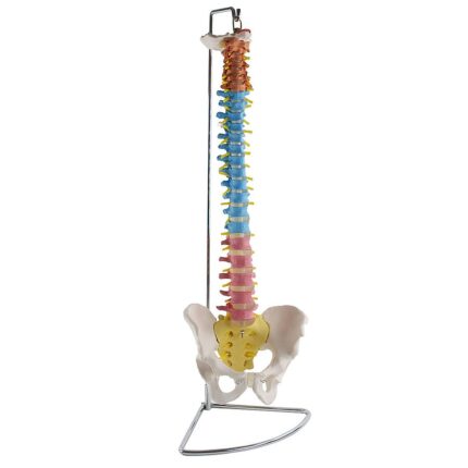
Spine model with color - H131267
The Spine Model with Color - H131267 is a life-size human spine. With color coding of the different spinal segments, the spine model is an excellent medical teaching aid.
The spine model consists of different vertebrae, which are grouped into the following sections:
-
- Cervical spine
-
- Thoracic spine
-
- Lumbar spine
-
- Sacrum and coccyx
-
-
- The cervical vertebra (C1-C7) in terracotta
- The thoracic vertebra (Th1-Th12) in light blue
- Male pelvis with symphysis
-
- In addition to the anatomical structures mentioned, the teaching spine model with pelvis also shows a herniated disc between L4 and L5. Thanks to the clear representation of the structure of the spine provided by this model, it is used as an undisputed teaching aid in universities and physiotherapy schools. The anatomical model is also ideal for use in doctors' offices and physiotherapy clinics for patient education. The teaching model of the spine and pelvis is provided on a fixed base, but can also be removed for better visualization.
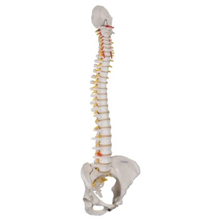
Spine model with pelvis 129993
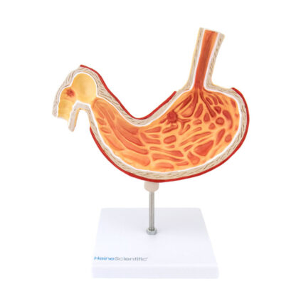
Stomach model with ulcer 139052
A model of a stomach ulcer from Heine Scientific represents the individual layers of the stomach as well as the connections from the esophagus to the stomach and from the stomach to the duodenum. In addition, ulcers appear in the stomach and duodenum. This stomach model is excellent for gastroenterological explanations to patients and as a teaching tool for medical students. Mounted on a stand.
Dimensions: 23 x 19 x 2 cm (H with base x W x D)
Base dimensions: 12 x 12 cm
Dimensions: 23 x 19 x 2 cm (H with base x W x D)
Base dimensions: 12 x 12 cm